Researchers Succeed in Reprogramming Skin Cells as Heart Cells
Findings are a Step Toward Techniques to Repair Damaged Hearts
 Researchers at University of Houston (UH) and Texas Heart Institute (THI) successfully
reprogrammed skin cells to function as early heart muscle cells. The results, published
in the August 7 issue of the Proceedings of the National Academy of Sciences, may lead to techniques to repair damage caused by heart attacks.
Researchers at University of Houston (UH) and Texas Heart Institute (THI) successfully
reprogrammed skin cells to function as early heart muscle cells. The results, published
in the August 7 issue of the Proceedings of the National Academy of Sciences, may lead to techniques to repair damage caused by heart attacks.
“During a heart attack, heart cells are injured and die, areas of the heart stiffen, and pump function is lessened,” said Robert Schwartz, the Cullen Distinguished University Professor in Biology at UH. “Since the heart doesn’t regenerate itself easily, we want to find ways to stimulate new heart cells and repair the injured heart.”
The team studied two proteins, ETS2 and MESP1, to see if they would work together to turn skin cells into heart cells. Schwartz, a UH developmental biologist, picked these two factors because they activate heart development in much simpler systems.
Skin Cells Convert to Heart Cells
“We used human skin cells for the study because they are really hard to convert,” said Schwartz, who also directs the Stem Cell Engineering Laboratory at THI. “Our thought was if ETS2 and MESP1 can turn skin cells into early, beating heart cells, then these factors will work in the human heart.”
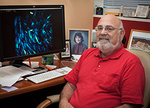
The research team added ETS2 and MESP1 proteins that enter skin cells in culture. The cells contained an internal marker that would turn red when the gene NKX2-5, an early cardiac factor, was activated.
“Within two days of adding the proteins, we saw that the cells were converting to immature heart muscle cells. We think that when introduced into a heart they would go on to mature as heart cells,” Schwartz said.
Heart Repair Experiments – Next Step
Schwartz wants to move forward with research to see if the technique can be used to repair heart damage. His lab is working on a method of inserting the proteins into the injured region of the heart using slow-release beads. The first studies would test the method in mouse and pig hearts.
“The idea is that by releasing these proteins slowly, we may convert cardiac fibroblasts or other cardiac cells into heart cells with the potential to repair the damage by replacing dead heart muscle,” he said. “We might be able to increase the number of heart cells and increase the vascular network.”
He is hopeful that the technique would have a therapeutic benefit of improved cardiac pumping and enhanced formation of blood vessels in the heart.
Schwartz collaborated with Vladimir N. Potaman of THI, and the study involved researchers and graduate students from UH, THI, Texas A&M Health Science Center in Houston, Baylor College of Medicine, and Sanford-Burnham Medical Research Institute in San Diego. The research was supported jointly through funds from the Cullen Foundation at UH and THI and through NIH grants.
- Kathy Major, College of Natural Sciences and Mathematics