Computational Medicine
Recent application of engineering quantification and analysis has skyrocketed in the field of medicine. Clinicians now rely on computational analysis for both clinical medical analyses, and basic science/clinical research. My research laboratory is conducting research in the following areas.
3D stereophotogrammetric imaging
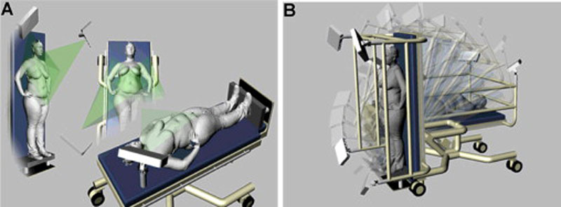
Our research developed an innovative 3D patent imaging mechanism to image patient in positions ranging from standing to supine. We mounted 3D imaging system on a bariatric tilt table to image the breasts at different angles between 0-90 degrees.
Visualization and breast metrics
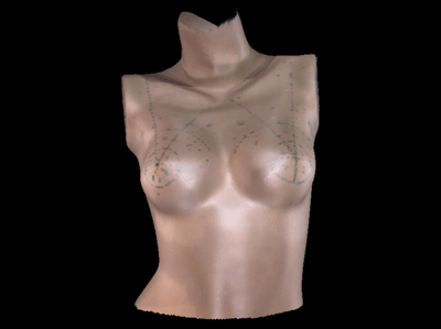
Interactive software for visualization, annotation and breast metric computation. The software enables basic 3D image processing such as rigid transformations and cropping. Manual annotation functionality is avaialbale for marking fiducial points. Breast metrics – distance, symmetry and volume measurements are computed using these fiducial points. We have utilized metrics computed using this software to quantify breast symmetry, quantifying breast cosmesis after conventional versus hypofractionated whole breast radiation in radiation oncology for breast conservation therapy in breast cancer treatment.
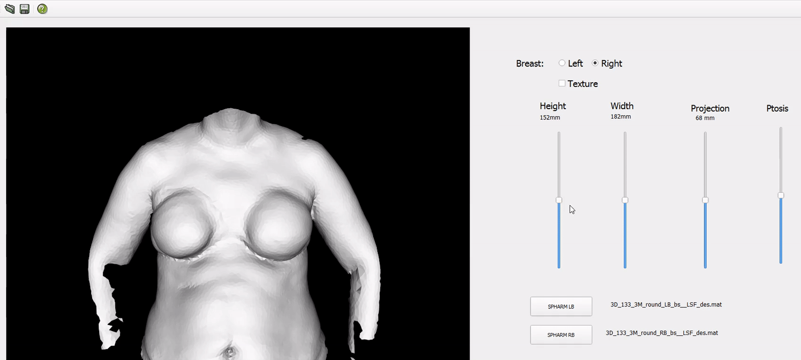
Breast shape modelling and simulation
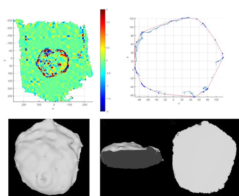
Intra-operative imaging for surgical planning
Spherical harmonics based computational modeling of breast shape for facilitating patient-physician communication during clinical consultation for breast reconstruction surgery. Our modeling approach leverages data-driven correlations of SPHARM shape descriptors with physical measurements of breast size (height and width), projection and ptosis to generate for an individual visualizations of breast shape based on their desired preferences. Controlled changes induced to SPHARM coefficients enables changes to the shape of the right breast shape occurring by adjusting, breast size, projection and ptosis.
Intra-operative imaging and analysis of mastectomy specimen for reconstruction surgery planning. We have developed imaging protocols to scan the mastectomy in the limited time between the mastectomy and reconstruction procedure. Our group has developed algorithms to process the specimen image. The algorithms include automated segmentation of the specimen from its background, computation of volume, width, height, depth and radii profiles.